
What We Do
We help research and medical laboratories upgrade their existing microscopes into powerful quantitative 3D imaging systems without replacing their current equipment.
Modular Microscope Upgrade
ZernIQ attaches directly to existing optical microscopes, turning them into compact Digital Holographic Microscopy systems with minimal workflow disruption.
Label-Free Imaging
Capture cellular structures without staining, labeling, or physical contact — enabling non-invasive observation for sensitive biological samples.
Quantitative 3D Reconstruction
Reconstruct holograms, phase data, intensity maps, and 1D/2D/3D profiles to gain deeper insight into microscopic structures.
Real-Time Cell Monitoring
Monitor subtle cellular changes and growth dynamics in real time, supporting live-cell compatible research and analysis.
Custom Integration & Calibration
We assess microscope compatibility, configure the module, optimize field of view and resolution, and support smooth lab integration.
Software & Long-Term Support
ZernIQ provides reconstruction and analysis tools, software updates, calibration, and future AI-assisted imaging modules.
Market Opportunity
Digital holographic microscopy is expanding rapidly, creating demand for affordable, modular, label-free 3D imaging solutions.
Global DHM market in 2025
Projected market by 2034
Expected CAGR
Netherlands module opportunity
TAM
Global digital holographic microscopy market
SAM
Serviceable market across Europe & the Netherlands
SOM
Initial launch opportunity focused on early-adopter laboratories
Based on internal market analysis from the ZernIQ business deck
Technology & Early Proof of Benefits
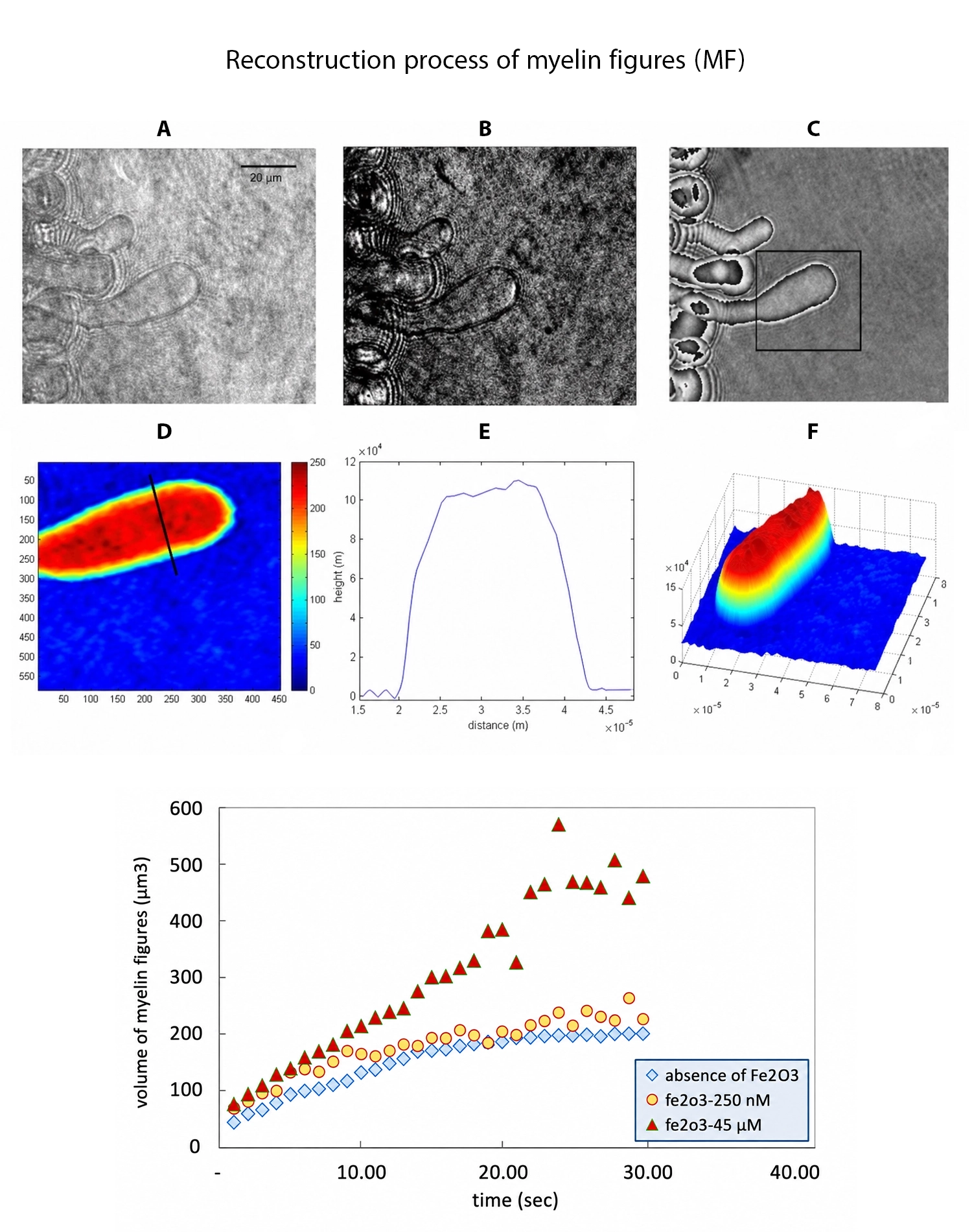
In our published study, “Detection of the Effect of Nanoparticles on Myelin Figures Growth Using a Compact Digital Holographic Microscope”, we investigated the effect of nanoparticles on the growth of myelin figures formed in phospholipid (lipid) systems.
Using a compact digital holographic microscope, we successfully monitored their growth dynamics in real time and quantitatively measured phase changes without staining or physical contact.